BREAST HEALTH SERIES, PART 1 OF 8
What a Mammogram Can Find, and What It Can Miss
By David Z. Schreier, MD, FACS | Breast and General Surgery | West Hills, CA
Breast cancer is the most common cancer in American women after skin cancer. The good news is that when it is found early, it is usually very treatable. That is the whole point of breast cancer screening: finding cancer before it causes any symptoms.
How Common Is Breast Cancer?
About 1 in 8 women in the United States will be diagnosed with breast cancer at some point in her life. For 2026, the American Cancer Society estimates:
- About 321,910 new cases of invasive breast cancer in women
- About 60,730 new cases of DCIS (ductal carcinoma in situ), a very early, non-invasive form of breast cancer
- About 42,140 deaths from breast cancer
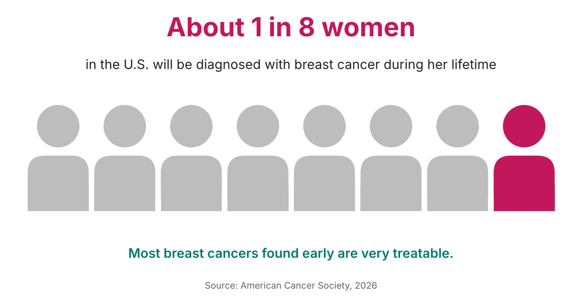
Risk goes up with age. The typical age at diagnosis is 63, but younger women can get breast cancer too. Men can also get it, although it is rare (about 1 out of every 100 cases).
There is real reason for hope. The breast cancer death rate has dropped about 44% since 1989, and there are now more than 4 million breast cancer survivors in the U.S. Earlier detection through screening and better treatment are a big part of that success.
When Should You Start Getting Mammograms?
For women at average risk, most major medical groups now agree: start at age 40.
- The U.S. Preventive Services Task Force recommends a mammogram every 2 years from age 40 to 74.
- The American College of Radiology and several other groups recommend every year starting at 40.
- Women at higher risk (for example, a strong family history, a BRCA gene change, or past radiation to the chest) may need to start earlier and may need extra tests, such as MRI. We’ll cover that in Part 2.
My recommendation: For women at average risk, I recommend a mammogram every 2 years starting at age 40, then every year starting at age 50. If you have other risk factors, we may start earlier or screen more often.
Not sure which schedule is right for you? Ask your doctor to review your personal risk. The American College of Radiology suggests every woman have a breast cancer risk check by age 25.
Where Does Ultrasound Fit In?
A breast ultrasound uses sound waves instead of X-rays, so there is no radiation. It does not replace a mammogram, because it can’t reliably see the tiny calcifications described below. But it is a very useful partner. Ultrasound is often used:
- To check a lump, or a spot seen on a mammogram, and tell whether it is a fluid-filled cyst or something solid
- As the first test for women under 30 with a lump, since younger breast tissue is usually dense
- During pregnancy or breastfeeding
- As an extra screening test for some women with dense breasts
- To guide a needle biopsy of a mass seen on ultrasound
What Is a Screening Mammogram?
A mammogram is a low-dose X-ray of the breast. Each breast is gently pressed between two plates for a few seconds while pictures are taken from two angles. The pressing can be uncomfortable, but it spreads the tissue out so the picture is clearer and the radiation dose stays low. 3D mammography (also called tomosynthesis) is now the standard at most of the imaging centers I work with. It takes pictures in thin slices, which can make small problems easier to see.
A screening mammogram is for women with no symptoms. A diagnostic mammogram is a more detailed exam used when there is a symptom, like a lump, or when something on a screening mammogram needs a closer look.
How a Mammogram Finds What You Can’t Feel: Calcifications
One of the most valuable things a mammogram can do is find calcifications. These are tiny specks of calcium in the breast tissue. They show up as small bright white dots on the X-ray. Many are smaller than a grain of sand, so there is no way to feel them during a breast exam.
Calcifications are very common, and most are harmless. They are not caused by drinking milk or taking calcium pills. They can come from aging, old injuries, or normal changes in the breast.
But certain patterns can be an early warning sign. When the specks are very small, packed closely together in a cluster, or lined up in a row or branching shape, they can be the first sign of DCIS or a small, early cancer. Finding cancer at this stage, often before it has formed any lump at all, is one of the biggest benefits of screening.

If your radiologist sees calcifications that look suspicious, the next step is usually close-up (magnified) mammogram pictures. If they still look concerning, a needle biopsy, referred to as a stereotactic biopsy, can be done. Because these spots can’t be felt, the doctor uses the mammogram machine to guide the needle to exactly the right place. This is a quick outpatient procedure, usually done with local numbing medicine.
Why a Lump You Feel May Not Show Up on a Mammogram
Here is something many people don’t know: a mammogram can be normal even when there is a cancer you can feel. Overall, screening mammograms miss about 1 in 8 breast cancers.
The main reason is breast density. Breasts are made of fatty tissue and dense tissue (glands and support tissue). On a mammogram, fat looks dark gray, while dense tissue looks white. Cancers also look white. So in a dense breast, a cancer can blend right in, like trying to spot a polar bear in a snowstorm.
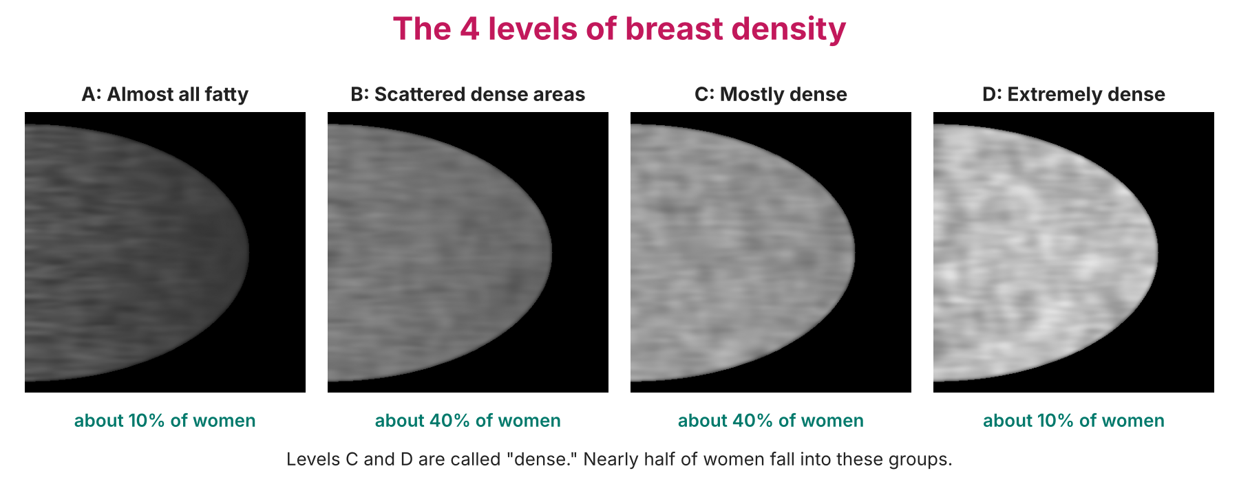
Nearly half of all women have dense breasts. Having dense breasts is normal, but it does make mammograms harder to read and somewhat raises breast cancer risk. Here in California, the law has required mammogram reports to tell women whether their breasts are dense since 2013, and a federal rule made this a requirement nationwide in September 2024. If they are, ask your doctor whether extra screening, such as ultrasound or MRI, makes sense for you.
Found a Lump? Don’t Let a Normal Mammogram Stop You
Screening tests are designed for people without symptoms. If you notice a change, you need a different approach. Call your doctor if you notice:
- A new lump or thick area in the breast or armpit
- Skin dimpling, puckering, redness, or skin that looks like an orange peel
- A nipple that turns inward when it didn’t before
- Bloody or clear fluid coming from one nipple on its own
- A change in the size or shape of one breast
Do this even if your last mammogram was normal. Your doctor will usually order a diagnostic mammogram and a breast ultrasound. A breast specialist or surgeon will then put the exam and imaging results together.
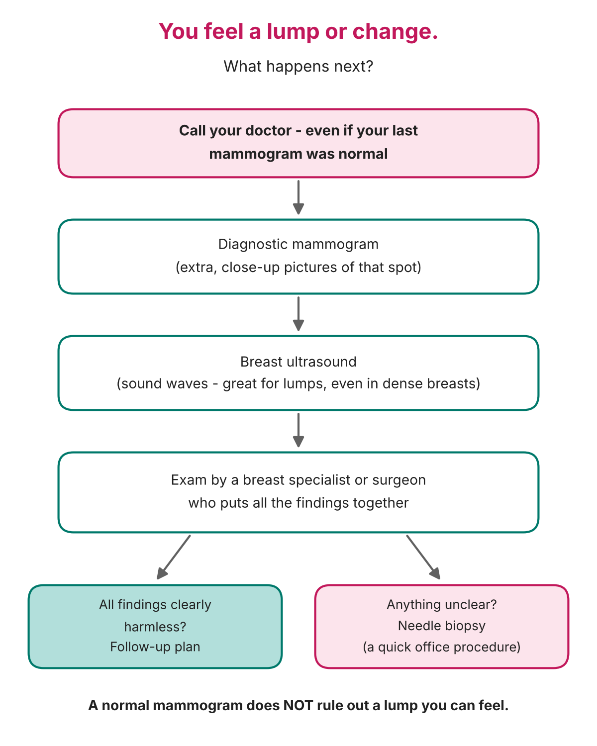
If anything is still unclear, a needle biopsy gives a definite answer. The good news: most breast lumps are not cancer. Many turn out to be cysts (fluid-filled sacs) or fibroadenomas (harmless, solid growths). But the only way to know for sure is to have it checked.
Called Back After a Mammogram? Try Not to Panic
Being called back for more pictures after a screening mammogram is common, especially after a first mammogram when there are no older pictures to compare. Fewer than 1 in 10 of the women who are called back turn out to have cancer. Usually, the extra pictures simply show normal overlapping tissue.
Key Takeaways
- About 1 in 8 U.S. women will get breast cancer in her lifetime, but early detection saves lives.
- Women at average risk should start mammograms at age 40.
- Mammograms can find tiny calcifications long before a lump can be felt.
- Dense breast tissue can hide cancer, so know your density.
- A lump or change you can feel always needs to be checked, even after a normal mammogram.
Coming Up Next
In Part 2, we’ll look at breast MRI: what it is, how it compares to a mammogram, its real advantages and disadvantages, and who should think about getting one.
Have a lump and/or unclear findings on your mammogram and ultrasound? Dr. Schreier’s office in West Hills can help. Contact us to schedule a visit.
Sources
- American Cancer Society. Key Statistics for Breast Cancer (2026). https://www.cancer.org/cancer/types/breast-cancer/key-statistics.html
- S. Preventive Services Task Force. Breast Cancer: Screening, Final Recommendation (2024). https://www.uspreventiveservicestaskforce.org/uspstf/recommendation/breast-cancer-screening
- American Cancer Society. Limitations of Mammograms. https://www.cancer.org/cancer/types/breast-cancer/screening-tests-and-early-detection/mammograms/limitations-of-mammograms.html
- American Cancer Society. Getting Called Back After a Mammogram. https://www.cancer.org/cancer/types/breast-cancer/screening-tests-and-early-detection/mammograms/getting-called-back-after-a-mammogram.html
- S. Food and Drug Administration. Mammography Quality Standards Act: Breast Density Reporting (2024). https://www.fda.gov/radiation-emitting-products/mammography-quality-standards-act-and-program
This article is for general education and is not a substitute for medical advice. Please talk with your own doctor about your personal screening needs.